
Fig. 4.
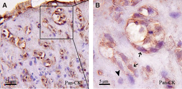
Immunohistochemical staining of pan-CK was detected in EMT-6/stromal cells reconstituted breast cancer. (A) pan-CK-positive EMT-6 cells dispersed in whole reconstituted breast cancer. (B) Higher magnification of the nest structures. EMT-6 (indicated by arrows) presented in reconstituted breast cancer tissue, and the stromal cells (indicated by arrowhead) accompanied by EMT-6 cells.