
Fig. 5.
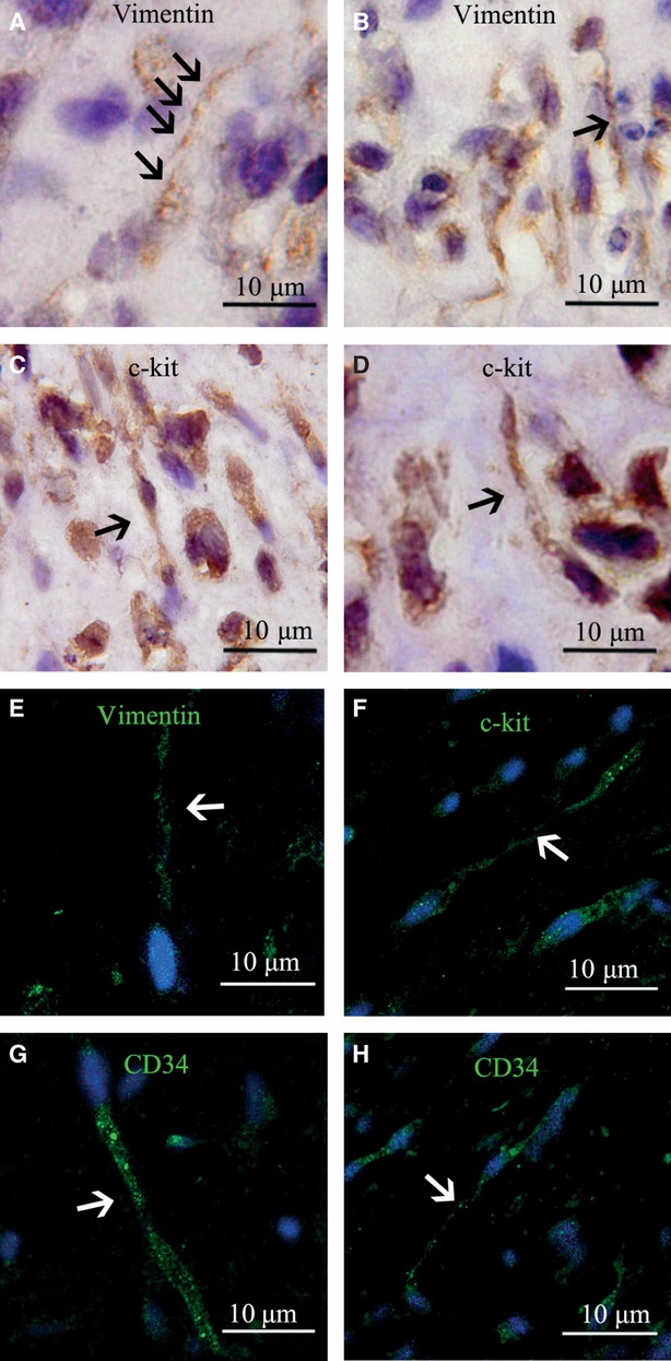
Immunohistochemical and immunofluorescent morphology of TCs in EMT-6/stromal cells reconstituted breast cancer tissue. (A–D) Immunohistochemical staining of TCs. (A) and (B) TCs were positive for vimentin with clear telopodes with dilations in A and with long prolongations in B (indicated by arrows, respectively). (C) and (D) TCs were positive for c-kit/CD117. (E–H) Immunofluorescent staining of TCs. The arrows indicate TCs. (E) Vimentin-positive TCs detected by immunofluorescence. (F) c-kit/CD117-positive TCs detected by immunofluorescence. (G) and (H) CD34-positive TCs detected by immunofluorescence.