
Fig. 6.
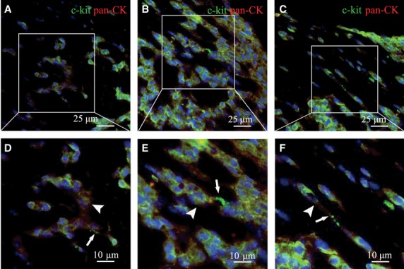
The spatial relationship between TCs and EMT-6 cells in EMT-6/stromal cells reconstituted breast cancer tissue was observed by double immunofluorescence staining. (A–C) c-kit/CD117+ cells was labelled with green colour and pan-CK+ cells was labelled with red colour. The nuclei were stained by Hoechst 33258. (D–F) Higher magnification of A, B and C. c-kit/CD117+ TCs distributed in the reconstituted breast cancer tissues (indicated by arrows), which are located close to pan-CK+ EMT-6 cells nest structure. EMT-6 cells were red stained and are indicated by arrowheads.