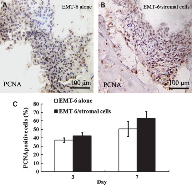
Fig. 8.

Comparison of the proliferation index by detecting the percentage of PCNA-positive cells in two groups of reconstituted breast cancer tissue. Immunohistochemical analysis of the expression of PCNA was evaluated in EMT-6 alone (A) and EMT-6/stromal cells reconstituted breast cancer tissue (B). Quantification of the proliferation index of PCNA-positive cells in EMT-6/stromal cells and EMT-6 alone reconstituted breast cancer tissue is shown in (C). Bars show the standard error of the mean.
