Fig. 1.

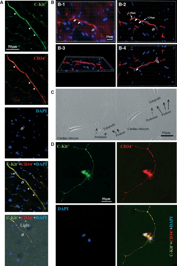
Identification of cardiac telocytes. (A) Double immuofluorescent staining for anti-c-kit (green) and CD34 (red) demonstrated c-kit+ and CD34+ cells with very small cell bodies (approximately 1:1 ratio of the cytoplasm to the nucleus; open arrow) and extremely thin prolongation in the interstitial space of cardiac myocytes. Some dilation was found in the thin prolongation of the c-kit+ and CD34+ cells (arrow). (B) 3D-recontruction of a representative cell further confirmed that the cell consisted of a small cell body with a nucleus (approximately 1:1 ratio of the cytoplasm to the nucleus; open arrow) and an extremely thin prolongation (diameter: approximately 1–2 μm; arrow). (B-1) representative Z-axis image. (B-2) 3-D reconstruction of B-1 with all Z-axis images. (B-3) 90° rotation of the X-axis of the images shown in B-2. (B-4) 180° rotation of the X-axis of the image shown in B-2. (C) Primary culture of isolated cardiac telocytes revealed that under phase-contrast microscopy, cardiac telocytes displayed piriform/spindle/triangular cell bodies and long, slender telopodia that contain alternations of the thick segments, i.e. podoms (arrow), and thin segments, i.e. podomers (dotted line arrow). (D) Cardiac telocytes with unique morphology are c-kit+ and CD34+. n = 3 for each group.
