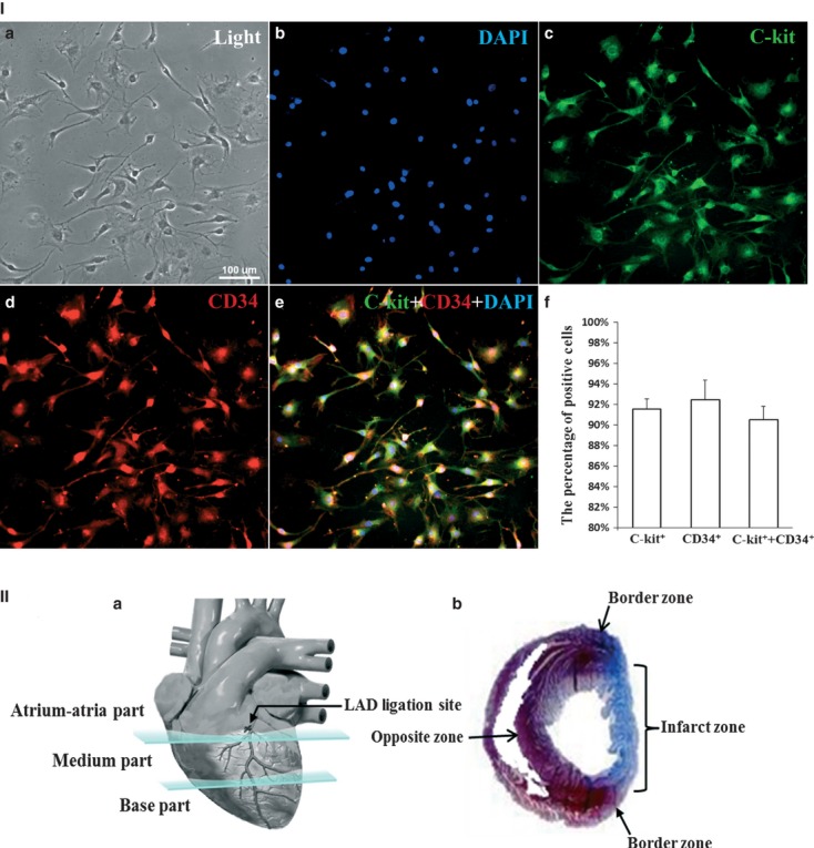
Fig. 2.

Purity of the isolated cardiac telocytes and schematic sections of the heart. (I) Double immunofluorescent staining for anti-c-kit and CD34 combined with cell counting demonstrated that approximately 91.5 ± 0.01% of the cells were c-kit positive, approximately 92.5 ± 0.02% of the cells were CD34 positive and approximately 90.5 ± 0.01% of the cells were c-kit and CD34 positive. a: light; b: DAPI; c: anti-c-kit (green); d: anti-CD34 (red); e: merged images from b, c and d. f: quantification of cells that were positive for c-Kit, CD34 and c-Kit and CD34 (n = 3). (II) a: schematic representation of the atrium-atria, medium and base parts of the heart; b: schematic representation of the infarcted zone, border zone and the zone opposite the infarcted zone.
