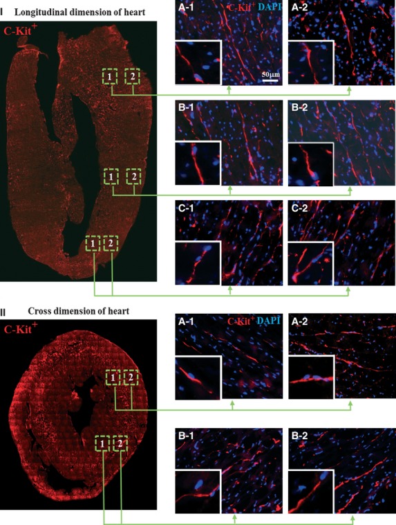
Fig. 3.

Distribution of cardiac telocytes in the myocardium. (I) Immuofluorescent staining for c-kit (red) revealed that many of the cardiac telocytes were distributed in the longitudinal dimension of the whole heart (A1-2, B1-2 and C1-2). (II) All of the cardiac telocytes were distributed within the cross direction (A1-2 and B1-2). Figures inset in I-A1-2, I-B1-2, I-C1-2, II-A1-2 and II-B1-2 contain images of cardiac telocytes at a higher magnification (n = 3).
