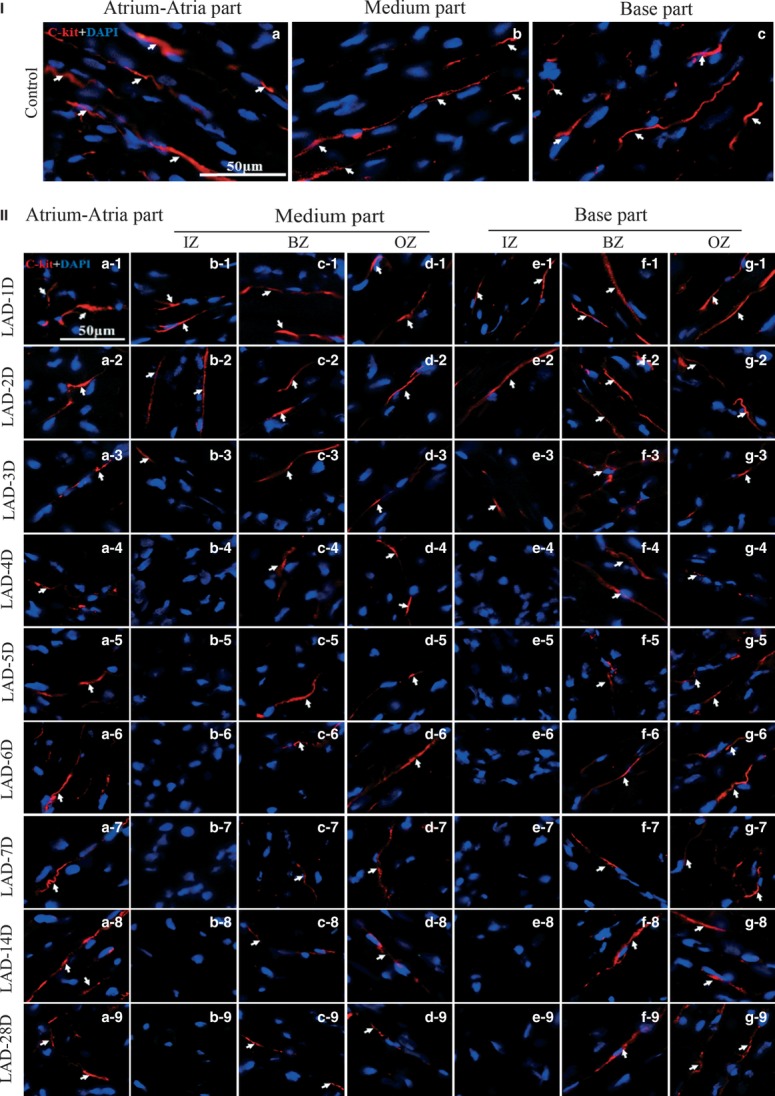
Fig. 4.

Comparison of cardiac telocytes in different segments of heart during MI. (I) Showing the cardiac telocyte density of table-1 in same region in the non-LAD ligated control of atrium-atria part (a), medium part (b) and base part (c). (II) Showing the change in cardiac telocyte density of table-1 for atrium-atria part of MI (a1-9); Showing the change in cardiac telocyte density of table-1 for infarcted zone of medium part of MI (b1-9); Showing the change in cardiac telocyte density of table-1 for border zone of medium part of MI (c1-9); Showing the change in cardiac telocyte density of table-1 for zone opposite the infarcted zone of medium part of MI (d1-9); Showing the change in cardiac telocyte density of table-1 for infarcted zone of base part of MI (e1-9); Showing the change in cardiac telocyte density of table-1 for border zone of base part of MI (f1-9); Showing the change in cardiac telocyte density of table-1 for zone opposite the infarcted zone of base part of MI (g1-9). IZ: infarcted zone. BZ: border zone. OZ: zone opposite the infarcted zone. n = 3.
