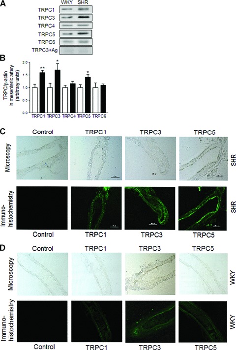
Fig 2.

Expression of TRPC1, TRPC3 and TRPC5 channels in mesenteric arterioles from SHR. Representative immunoblottings (A) and summary data (B) of TRPC1, TRPC3, TRPC4, TRPC5,TRPC6 channels and TRPC3 antibody with its respective antigenic peptide the protein expression in mesenteric arterioles from WKY (open bars) and SHR (filled bars). Data are mean ± S.E.M. of n= 4 independent experiments. *P < 0.05 or **P < 0.01 by two-tailed Student’s t-test. (C) Light microscopy of mesenteric arterioles from SHR (upper panels) and immunohistochemistry (lower panels) of TRPC1, TRPC3 and TRPC5 protein expression in mesenteric arterioles from SHR. (D) Light microscopy of mesenteric arterioles from WKY (upper panels) and immunohistochemistry (lower panels) of TRPC1, TRPC3 and TRPC5 protein expression in mesenteric arterioles from WKY. Immunohistochemistry using specific antibodies labelled with green shows TRPC1, TRPC3 and TRPC5 protein expression in mesenteric arterioles. Control indicates immunohistochemistry after pre-incubation of the primary TRPC3 antibody with antigenic peptide for 12 hrs at 4°C. Bar denotes 200 μm. (43.0 ± 15%; P= n.s.; n= 6, Fig. 4D). In the presence of anti-TRPC5 antibodies the norepinephrine-induced vasomotion was not significantly affected (45.0 ± 11%; P= n.s. Fig. 4E). In the presence of anti-TRPC1 plus anti-TRPC3 antibodies the norepinephrine-induced vasomotion was significantly reduced to 29.0 ± 8% (P < 0.05; n= 6, Fig. 4F). Furthermore, pre-incubation of mesenteric arterioles with anti-β-actin antibodies did not significantly affect norepinephrine-induced vasomotion when compared to control conditions (59.8 ± 15%, P= n.s.; n= 6, Fig. 4G). Incorporation of TRPC antibodies into mesenteric arteries by pinocytosis was facilitated by changing the bathing solution to a hypotonic solution according to recent literature [17].
