
Fig 6.
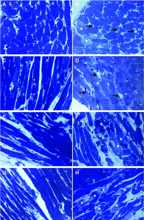
Light microscopy of toluidine blue stained semi-fine sections of Epon-embedded tissue. a – control, b – IR, c – Resveratrol, d – Resveratrol + 3 MA, e –γ-Tocotrienol, f –γ-Tocotrienol + 3 MA, g – Resveratrol +γ-Tocotrienol, h – Resveratrol +γ-Tocotrienol + 3 MA. Cardiac tissue proves to have almost normal structure on light microscopy in control (a), Resveratrol (c), γ-Tocotrienol (e) and Resveratrol +γ-Tocotrienol (g) treated samples. Oncotic changes of cardiomyocytes characterized by contraction band necrosis (*) and vacuolar degeneration (arrows) are visible in IR (b) and 3MA supplementary treatment (d, f, h).