Fig. 1.

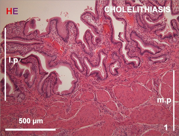
Cross section of the gallbladder wall from the cholelithiatic group showing mild infiltration with inflammatory cells and a thickened muscular layer. H&E staining. l.p., lamina propria; m.p., muscularis propria.

Cross section of the gallbladder wall from the cholelithiatic group showing mild infiltration with inflammatory cells and a thickened muscular layer. H&E staining. l.p., lamina propria; m.p., muscularis propria.