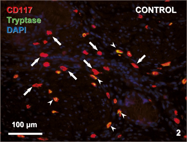
Fig. 2.

Cross section of the gallbladder wall of a control patient stained for CD117 (red) and tryptase (green). The nuclei are counterstained with DAPI (blue). CD117-positive/tryptase-negative TCs (arrows) and CD117-positive/tryptase-positive mast cells (arrowheads) are indicated.
