
Fig 6.
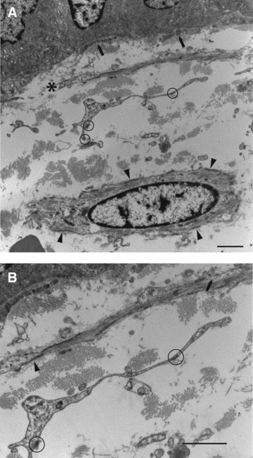
(A) (top): Cell processes and a nucleated cell profile. Closest to the epithelium (∼80 nm) are slender subepithelial stromal cell processes of indeterminate nature (arrows). Next, in terms of distance from the epithelium, is a slender cell process (*) showing smooth-muscle features (many myofilaments and multiple attachment plaques). This is ∼1.3 μm from the basal lamina at its nearest and so by definition is subepithelial. Then, there is a cell process ∼5 μm away from the basal lamina, and is also therefore, by definition, subepithelial: it is of fibroblastic appearance – no lamina or filaments, but focal adhesions (circles). Finally, at the bottom of the figure is an interstitial stromal cell, ∼10 μm distant from the basal lamina, with unambiguous smooth-muscle features (prominent myofilaments and attachment plaques, arrowheads). (B) (bottom): Details of Figure 6A showing lamina (arrowhead), myofilaments and plasmalemmal caveolae (arrow) of the smooth-muscle type subepithelial stromal cell and the focal adhesions (circles) of the fibroblastic subepithelial stromal cell process. Normal colon, case 15. Bar, 1 μm.