
Fig 13.
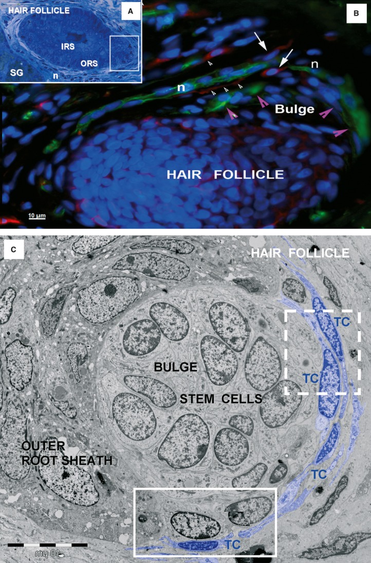
Telocytes around human hair follicle and adjacent sebaceous gland. (A) Light microscopy image of toluidine-blue coloured, thin section shows a hair follicle and an adjacent sebaceous gland (SG). IRS: inner root sheath; ORS: outer root sheath; n: perifollicular nerve fibres. (B) Epifluorescence microscopy: double labelling shows nestin positive cells (green), stem cells from the bulge area of hair follicle (magenta arrowheads) and perifollicular nerve fibres (N), surrounded by CD117 positive TCs (red, white arrows) with long telopodes (white arrowhead). Nuclei are counterstained with DAPI (blue). Original magnification 400×. (C) TEM of the boxed area in (A) shows a cluster of stem cells in the outer root sheath of a hair follicle. The stem cells are bordered by telocytes (TC) with telopodes, and other cells from the outer root sheath. Boxed areas are enlarged in Fig. 14.