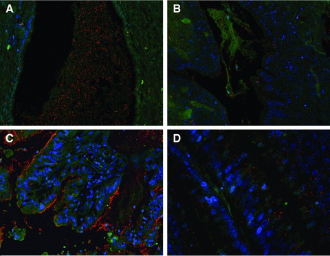
2 (A, B).

Mucinous ovarian carcinoma expressing Tn-MUC6 (A) predominantly at the extracellular mucin pools and SLea-MUC5AC glycoform (B) at cell cytoplasm and extracellular mucus. PLA images at 200x magnification. (C) Mucinous gastric carcinoma expressing STn-MUC1. PLA signals are seen at cell cytoplasm, apical membrane and extracellular mucus, at 200x magnification. (D) Mucinous colon carcinoma expressing Tn-MUC5AC at a perinuclear, Golgi-like, location, at 400x magnification. In this case, PLA signals suggest that the secreted MUC5AC had further processing of the Tn antigen which explains negativity by PLA at secreted mucus.
