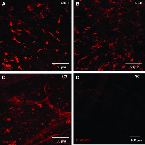
Fig 4.

Confocal imaging of vimentin-positive cells in lamina propria. (A, B) Whole mount, flat sheet preparations of sham bladder mucosa were labelled with anti-vimentin and imaged with confocal microscopy. All images are reconstructions of optical sections. Networks of vimentin-positive cells were found in the lamina propria. Vimentin-positive cells had bipolar or highly branched stellate morphology. (C) Mucosal tissues from SCI animals also had vimentin-positive cells however, the networks were apparently less well developed than the sham operated controls and cells had a compromised, non-branched morphology. (D) Tissues labelled with only the secondary antibody (primary antibody omitted) had minimal staining.
