Fig 6.

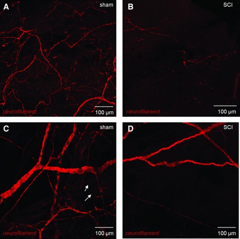
Confocal imaging of bladder neural networks. (A) Whole mount, flat sheet preparations of mucosa from sham bladders were labelled with anti-neurofilament and imaged with confocal microscopy. All images are reconstructions of optical sections. A neural plexus was observed in these sections comprising larger nerves, which bifurcated to increasingly smaller fibres. (B) In SCI animals, the neural plexus was significantly disrupted with an apparent reduction in nerves. (C) The detrusor region of sham control bladders was densely innervated with large nerve trunks subdividing into increasingly smaller nerves, which became varicosities (arrows) within the smooth muscle bundles. Mucosal tissues from SCI animals also had strikingly compromised neuronal distribution with only occasional patches of immunopositive nerves. (D) In SCI detrusor, the innervation was comparatively sparse with only patchy areas of nerves visible.
