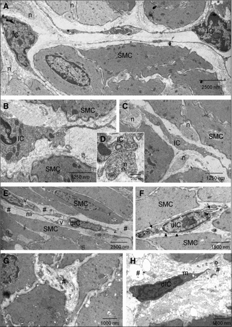
Fig 9.

Transmission electron micrographs of detrusor IC in sham and SCI bladders (A–C) Cells with ultrastructural characteristics of IC were present in sham detrusors. These had cytoplasmic branches and were close to smooth muscle cells (SMC). Nerve (n) varicosities were abundant and were in close contact with both SMC and IC. (D) Magnification of the boxed area in B showing synapse-like contact between an IC and a nerve ending. (E–H) After SCI, damaged IC (dIC) were present with evidence of damage to branched processes and cell–cell contacts. dIC typically had cytoplasmic vacuolisation (v), swollen endoplasmic reticulum (arrows), apparent collapse of the cytoskeleton in the perinuclear area (arrowheads) and some swollen mitochondria (m). Areas of cellular debris were abundant (#). SMC exhibited a lesser degree of degradation.
