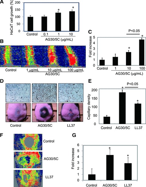
Fig 5.

Evaluation of the angiogenic effects of AG30/5C in a wound-healing model. (A) Cell growth of HaCaT cells in the presence of AG30/5C (0, 0.1, 1 and 10 μg/ml). The quantification is presented as a percent increase. N = 8 per group and duplicated. (B, C) Blood flow was analysed by Laser Doppler Image in mouse tails three days after induction of full thickness injuries with topical application of AG30/5C (0, 1, 10 and 100 μg/ml). Low or no perfusion is displayed as dark blue, whereas the highest perfusion interval is displayed as red; the quantification is presented as a fold increase. N = 3–5 per group and duplicated. *P < 0.05 versus control. (D, E) Evaluation of wound healing in a diabetic mouse model treated with saline (control), AG30/5C or LL37. (D) The upper panel shows representative immunostains using anti-CD31 (PECAM) antibody (brown). The lower panel shows representative pictures after wound closure (day 24). Hair growth was observed around the edge of wound. (E) Capillary densities were quantified by cross section of ischaemic tissues immunostained with anti-CD31 (PECAM) antibody. N = 6–10 per group and duplicated. *P < 0.05 versus control. (F, G) Blood flow was analysed by LDI in mouse wound 3 days after operation with topical application of AG30/5C (100 μg/ml). Low or no perfusion is displayed as grey or dark blue, whereas the highest perfusion interval is displayed as yellow or red; the quantification is presented as a fold increase. N = 5–6 per group and duplicated. *P < 0.05 versus control.
