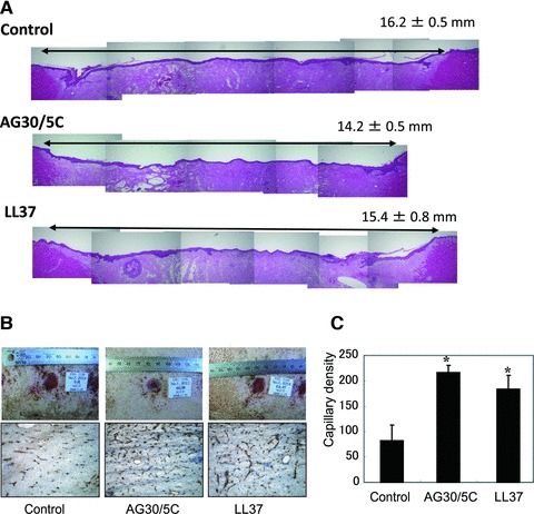
Fig 7.

Evaluation of the effects of AG30/5C on a porcine wound-healing model. (A) Representative pictures of haematoxylin and eosin staining on full-thickness section with restored epithelium, treated with saline (control), AG30/5C or LL37 (100 μg/ml, respectively). In the upper right corner, means ± S.E.M. (mm) were shown. (B) The upper panel shows representative pictures of wound healing in each group (day 9). The lower panel shows representative immunostains using anti-vWF antibody (brown). (C) Capillary densities were quantified by cross section of ischaemic tissue immunostained with anti-vWF antibody. *P < 0.05 versus control. N = 6 per group and duplicated.
