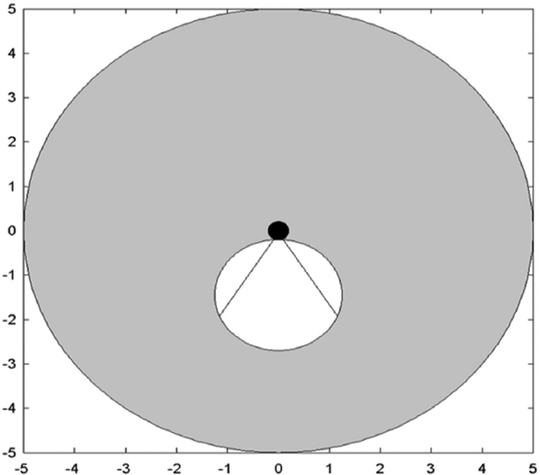
Figure 1.
Schematic of idealized dual-mode IVUS operation. Radial imaging field of view (gray) for circular phased array. White circle shows boundaries of the tumor. Hyperthermia beam (straight lines show full width at half maximum) from catheter (black) directed at tumor. Scale in centimeter.
IVUS = intravascular ultrasound.

