
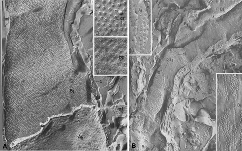
1.
Endothelium of continuous (A) and fenestrated (B) types as seen by freeze fracture. (A) The P face of the abluminal membrane of a heart endothelial cell shows the numerous caveolae. These are organized in linear arrays, better seen in the higher magnification images (insets). (B) Two endothelial cells of a jejunal capillary show the disposition of fenestrae in sieve plates. Higher magnification insets compare the linear disposition of caveolae (left) and fenestrae (right), suggesting attachment to cytoskeletal elements. Reproduced from reference [12], with permission.