6.

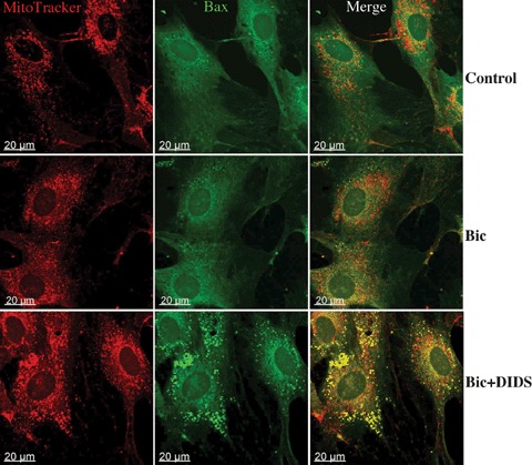
Immunostaining analysis of Bax location in EC (similar experimental conditions as in Fig.3). Mitochondria were labeled with Mitotracker (red) and Bax with specific antibodies (green). Yellow spots in merge images represent the colocalization of Bax with mitochondria. Data are representative from three independent experiments with similar results.
