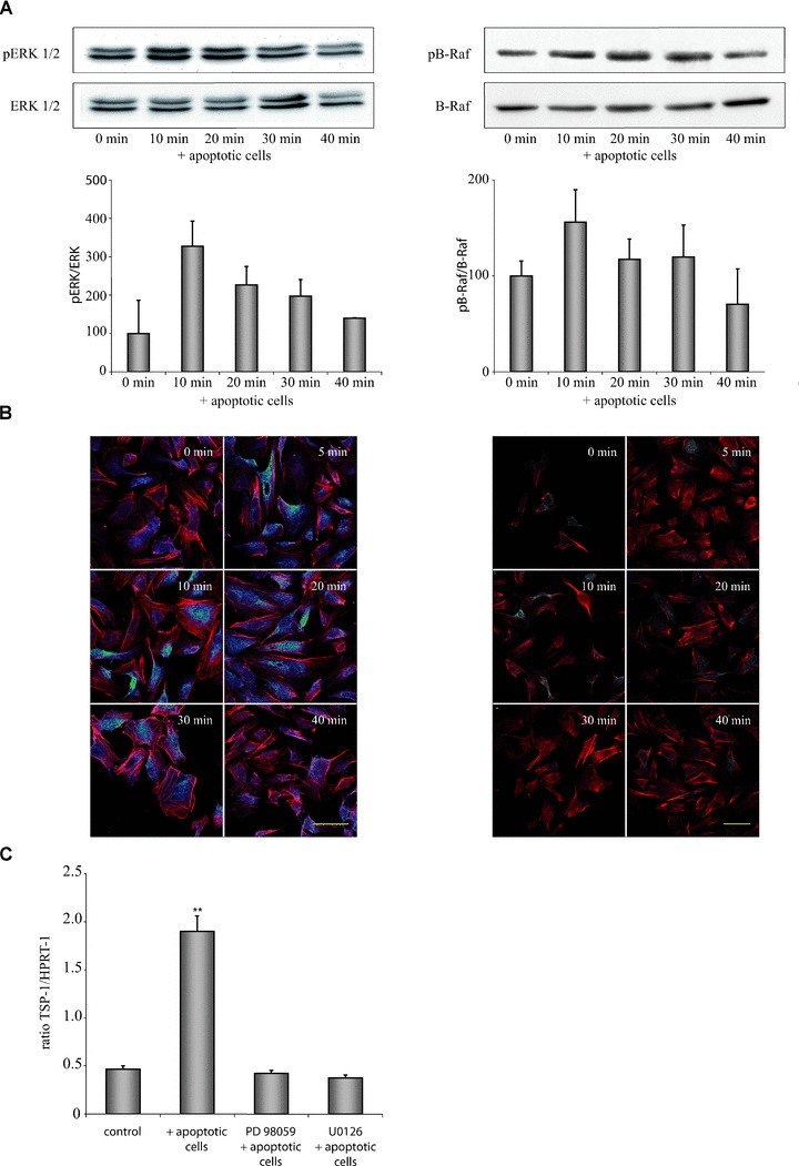
Fig 2.

Apoptotic cell-induced expression of TSP-1 in HUVEC is mediated by the B-Raf/MEK/ERK pathway. (A) Exposure of HUVEC to apoptotic eEND2 cells led to a rapid activation of ERK1/2 (left) and B-Raf (right). Changes in phosphorylation were determined by calculation of the ratio of phosphorylated to unphosphorylated ERK1/2 or B-Raf protein, respectively (n= 6). (B) Immunocytochemical staining of pERK1/2 and pB-Raf in HUVEC exposed to apoptotic eEND2 cells. Cells were counterstained with phalloidin-Alexa546 and analysed by confocal microscopy. Shown are representative micrographs of three independent experiments. Scale bars represent 50 μm. (C) Inhibition of ERK phosphorylation by U0126 or PD98059 circumvented apoptotic cell-induced enhanced TSP-1 expression in HUVEC as assessed by real-time qPCR measurement (n= 4; **P < 0.001).
