Fig 3.

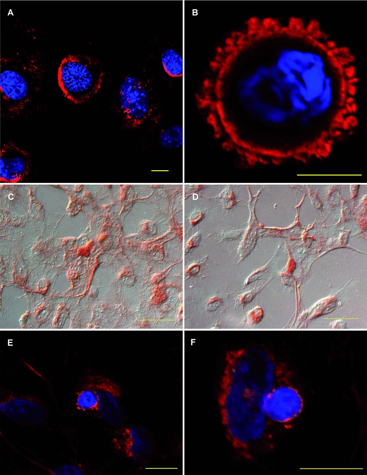
HUVEC-derived TSP-1 is bound by apoptotic murine eEND2 cells. (A, B) Apoptotic murine eEND2 cells were incubated with conditioned supernatant of HUVEC exposed to apoptotic cells and stained for human TSP-1. Shown are representative confocal images of four independent experiments. The nucleus was counterstained with DAPI. Scale bar represents 10 μm. (C, D) HUVEC were exposed to apoptotic eEND2 cells for 30 min. Cells were counterstained with haematoxylin and eosin and analysed by conventional microscopy. (E, F) HUVEC were seeded onto μ-slides and exposed to apoptotic eEND2 cells for 30 min. Cells were stained for human TSP-1 and applied to confocal microscopy. The nucleus was counterstained with DAPI. Scale bars represent 50 μm.
