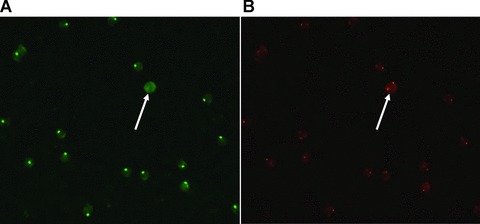
Fig 3.

K562 cell (female origin) spiked onto slide containing male lymphocytes. Show Target cell of selection #3.2. (A) Shows the result of FISH using a Y-chromosome specific probe (green) and (B) shows the result of FISH using an X-chromosome specific probe (red). Hybridization was performed twice, reversing the colours of the chromosomes; the figure showing the last FISH. After the first hybridization Y- and X-chromosomes were red and green, respectively. Colours were reversed in a second hybridization (shown in the figure) to eliminate false positive cells.
