Fig 3.

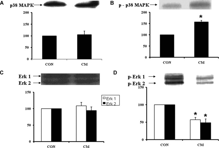
Western blot analysis of p38 MAPK (A), phosphorylated p38 MAPK (B), Erk1/2 (C) and phosphorylated Erk 1/2 (D) in control (CON) and cardiomyopathic (CM) hamster hearts. *P < 0.05 versus control; (n= 6).

Western blot analysis of p38 MAPK (A), phosphorylated p38 MAPK (B), Erk1/2 (C) and phosphorylated Erk 1/2 (D) in control (CON) and cardiomyopathic (CM) hamster hearts. *P < 0.05 versus control; (n= 6).