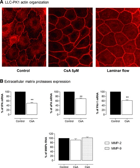
Fig 1.

CsA-induced reorganization of actin filaments and decrease in extracellular matrix proteases in proximal tubular cells. (A) LLC-PK1 cells were exposed to CsA 5 μM or laminar flow (1.65 mm/sec.) for 24 hrs then stained with fluorescein-labelled phalloidin and analysed by immunofluorescence microscopy (40×). (B) mRNA of tPA, urokinase, PAI-1 and metalloproteinases 2 and 9 (MMP2, MMP9) were quantified by qPCR. Pictures highlight modification in global cell’s shape, stiffening of the lateral actin network, and a decrease in tPA, urokinase and PAI-1 expression under CsA conditions.
