
Fig 1.
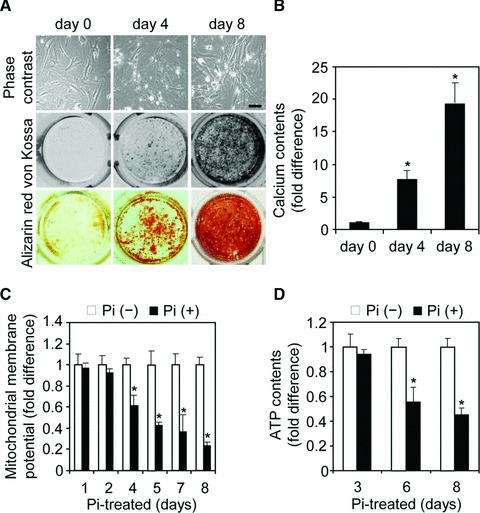
Impairment of mitochondrial function during Pi-induced VSMC calcification. (A, B) Pi-induced VSMC calcification. VSMCs (2 × 10⁴ cells/well) were seeded in a 48-well culture plate and cultured until confluence. After cells were treated with 2.6 mM Pi for the indicated times, calcium deposits were visualized by staining with von Kossa (brown or black) and alizarin red S (red) (A), and calcium content was measured (B). A representative experiment is presented in von Kossa and alizarin red S analysis. Scale bar in (A) indicates 200 μm. (C, D) Declines in mitochondrial membrane potential and intracellular ATP levels during Pi-induced VSMC calcification. VSMCs (1 × 10⁵ cells/well in a six-well culture plate) were grown to confluence and then treated with Pi for the indicated times. Mitochondrial membrane potentials (C) and intracellular ATP levels (D) were measured by flow cytometric analysis of rhodamine 123 fluorescence and by a bioluminescence assay, respectively. Data are expressed as the mean ± S.D. (n= 3). *P < 0.01 versus Pi-untreated control.