
Fig 5.
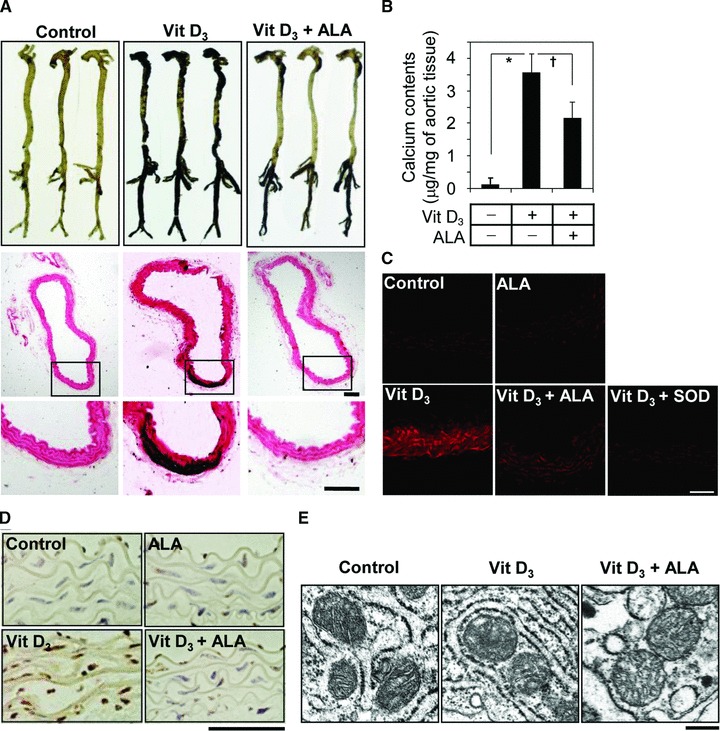
ALA protects vitamin D3-induced aortic calcification in mice. (A) von Kossa staining for mineral deposition in aorta. Whole aortas were dissected from naïve and challenged mice and stained with von Kossa (upper). n= 3. Cross-sectioned specimens of the middle part of thoracic aorta were stained with von Kossa, counterstained with nuclear fast red, and visualized by light microscopy (middle). The boxed area of middle panel is enlarged in lower panel. (B) Calcium content of whole aortas was measured and normalized by semi-wet dried weight of aortic tissues. Data are expressed as the mean ± S.D. (n= 3). (C) To detect superoxide anion in aortic tissues, cryosections of aortas freshly harvested at 6 days after final vitamin D3 injection were stained with DHE and the fluorescence images were captured under confocal microscopy. Peg-SOD was used to verify that the fluorescent signals are specific for superoxide anion. (D) To detect apoptotic cells in aortic tissues, aortas were sectioned, stained with TUNEL, and visualized under light microscopy. (E) Electron microscopic images of mitochondria present on aortic tissues. A representative experiment in (A) and (C–E) is presented. Scale bars in (A) and (C–E) indicate 100, 50, 200 and 500 nm, respectively. *P < 0.01; †P < 0.05.