
Fig 1.
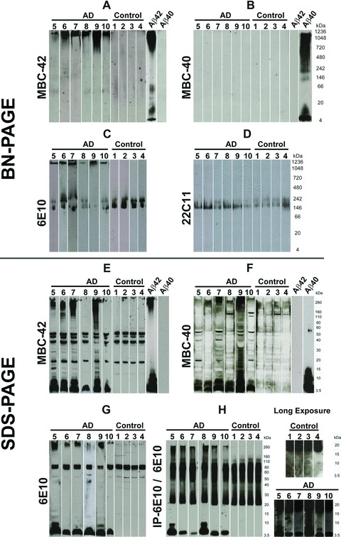
Western blot analysis of sucrose soluble proteins from AD and control brains after BN-PAGE (A–D) and after SDS-PAGE (E–H). All BN-PAGE blots were developed 2–3 hrs for chemiluminescence exposure. SDS-PAGE blots were exposed for 2–5 min. (A) The protein lysates from AD brains (cases no. 5–10) in BN-PAGE showed a high-molecular weight anti-Aβ42-positive smear >1000 kD. Such smears were not observed in controls (cases no. 1–4). Synthetic Aβ42 and Aβ40 were loaded as positive and negative controls, respectively. In Aβ42 preparations, long chemiluminescence exposure led to the detection of additional dimer and ∼50 kD bands that were not observed after 2–5 min. exposures, as shown in Figure 3C. (B) The Aβ42-positive material seen in (A) was not detectable in AD (cases no. 5–10) or in the controls (cases no. 1–4) in the native gel blotted with anti-Aβ40 antibodies. Synthetic Aβ42 and Aβ40 were loaded as positive and negative controls, respectively. After 3 hrs of chemiluminescence exposure, synthetic Aβ40 blots display a dimer band at ∼10 kD in addition to the monomer band and the hiMWAβ smear already detected with shorter exposure times as depicted in Figure 3C. (C) The anti-Aβ1–17 antibody also detected the high-molecular weight protein aggregates >1000 kD in the area of stacking gel in the protein lysate from AD brains (cases no. 5–10), which was not detectable in control brains (cases no. 1–4). In addition, anti-Aβ1–17 also showed APP bands in AD and control cases (140–240 kD). (D) The APP-positive bands were confirmed with an antibody directed against N-terminal epitope of APP (22C11) in control (cases no. 1–4) and AD cases (cases no. 5–10). (E)–(G) SDS-PAGE analysis of AD brain protein lysates from cases no. 5–10 exhibited Aβ monomer and dimer bands with MBC-42 (E), MBC-40 (F) and anti-Aβ1–17 (G) that were not detected in control brains (cases no. 1–4). The MBC-42-dimer (E) and 6E10-dimer bands (G) were not seen in all AD cases, whereas anti-Aβ40 consistently detected dimer bands (F). A high-molecular smear was found in most AD cases with all three antibodies directed against Aβ. Interestingly, cases 6 and 10 exhibited nearly no SDS-stable hiMWAβ42 aggregates (E), whereas both cases showed high-molecular anti-Aβ42-positive material in the BN-PAGE (A). (H) With the help of anti-Aβ1–17 (6E10)-immunoprecipitation, monomer and dimer bands as well as loMWAβ (4–20 kD) smears and hiMWAβ (>160 kD) smears were visible in SDS-PAGE of AD brain lysates (cases no. 5–10) but not in those of controls (cases no. 1–4). The detection of the loMWAβ oligomers required chemiluminescence exposure for 3 hrs (i.e. long exposure times).