Fig 2.

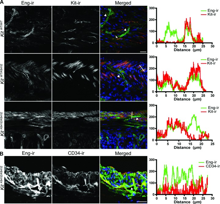
Eng expression in endothelium and in Kit+ ICC in WT and KitK641E antrum. (A) Eng-ir (Alexa 488, green) was observed in Kit-ir ICC (NL559, red) (indicated by arrow) in KitWT/WT antrum. The higher abundance of Kit+ cells in KitK641E heterozygotes and homozygotes made Eng-ir even more evident. Robust Eng-ir was also consistently found in the endothelium of blood vessels (indicated by arrowhead) in all genotypes. (B) Example of Eng-ir (Alexa 488, green) and CD34-ir (NL559, red) in P14 KitK641E/K641E antrum. Blood vessels (indicated by arrowhead) were decorated by both CD34-ir and Eng-ir, although CD34− cells of the hyperplasic longitudinal muscle layer exhibited also Eng-ir. Figures oriented with the serosa facing up and mucosa down. Scale bar: 20 μm. Intensity plots for the green and red fluorochromes were measured along the white line drawn on the respective merged image.
