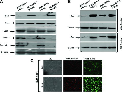
Fig 5.

(A) Western blot analysis demonstrates induction of Bax and inhibition of XIAP, Mcl-1 and surviving by the induction of APR-1 protein without influencing the basal expression level of Bak. β-actin was used as internal control for loading and transfer. (B) Western blot analysis of Bax protein in both mitochondrial and ER fractions. The purity of both mitochondrial and ER fractions was assessed by the detection of Tom20 (mitochondrial marker) and Bap31 (ER marker) in the corresponding fractions. (C) Intracellular Ca2+ levels in BLM-APR-1 cells before and after the addition of Dox (10 ng/ml) to the culture medium for 24 hrs as assessed by staining with Ca2+-sensitive dye Fluo3-AM and Mitotracker red. Differential interference contrast images (DIC) are shown. Data are representative of three independent experiments.
