
Fig 4.
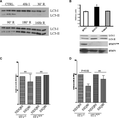
(A) Simulated I/R of isolated primary cardiac myocytes showed LC3-II protein levels being induced from 90 min. reperfusion. Western blot analysis at various time-points and immunoblotting with the indicated antibodies (B) IFN-γ-STAT1 activation reduced and epigallocatechin-3-gallate-reduce STAT1 activation enhanced conversion of LC3-I to II following simulated I/R of isolated primary cardiac myocytes. Primary cardiac myocytes were treated with either IFN-γ (50 ng/ml) or epigallocatechin-3-gallate (EGCG 50 ng/ml) and exposed to simulated I/R and cells harvested after 180 min. and processed for Western blot with the indicated antibodies (lower panel). Upper panel shows densitometric analysis of LC3-I over LC3-II ratio. (C) Enhanced autophagic flux in STAT1-deficient (ST1−/−) cells following serum starvation (SS) using the ployQ80 luciferase reporter assay. Wt (ST1+/+ or STAT1-deficient (ST1−/−) MEF cells were transfected with polyQ80luc and subjected to serum starvation (SSQ80) or control conditions (NDQ80). At 6 hrs of nutrient deprivation, no significant difference in autophagic turnover of the polyQ80 aggregates was seen. (D) After 18 hrs of nutrient deprivation the ST1−/− MEFs showed nearly 59% decrease in polyQ80 luciferase activity, whereas the ST1+/+ MEFs showed no statistically significant decrease in polyQ80 luciferase activity The data above show pooled data from three experiments with the NDQ80 set at 100% and the data analysed using a paired Student’s t-test to compare NDQ80 versus SSQ80.