
Fig 1.
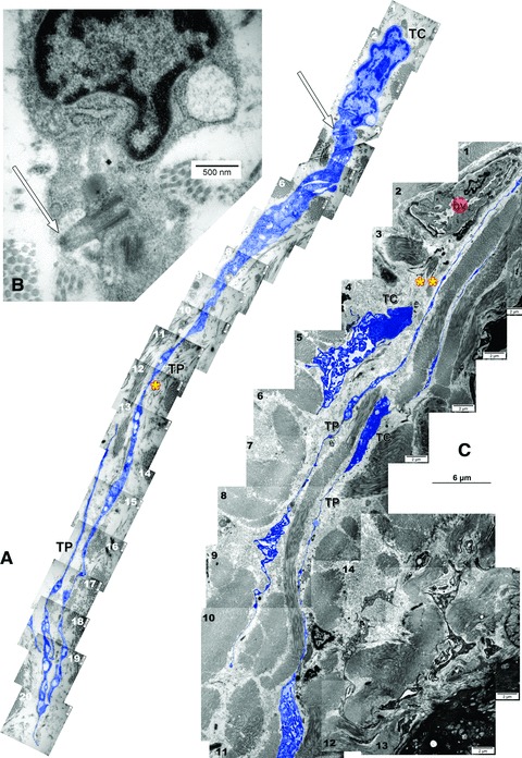
Ultrathin sections of rat trachea. Two-dimensional sequenced concatenation from 20 (A) and 16 (C) serial electron micrographs depicting telocytes (TC) with characteristic long and slender prolongations (TP: telopodes): (A) *= 44 micrometers; (B) **= 45 micrometers. In (B) is detailed a primary cilium (arrow) of the telocyte in (A). bv: blood vessel. Telocytes and telopodes were digitally coloured blue.