3.

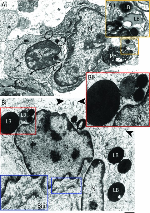
Activated heart macrophages triggered by the acute Trypanosoma cruzi infection. A clear interaction between an activated macrophage and a lymphocyte is observed in (Ai). Note areas of cell-to-cell attachments (circles). A phagolysosome is seen within the macrophage cytoplasm in close apposition to lipid bodies (LB) (box). (Aii) Higher magnification of the boxed area shows LBs with different electron densities and sizes. (Bi) Other features of macrophage activation include increase of both surface projections (arrowheads) and LB numbers and prominent Golgi complex profiles. Boxed areas show details of LBs and Golgi in higher magnification in Bii and Biii, respectively. Heart samples were processed for transmission electron microscopy at day 12 of infection. N, nucleus; My, myocardium. Scale bar, 600 nm (Ai); 400 nm (Aii); 750 nm (Bi) and 500 nm (Bii and Biii).
