
Fig. 3. NK cells express pro-resolving receptors and respond to LXA4.
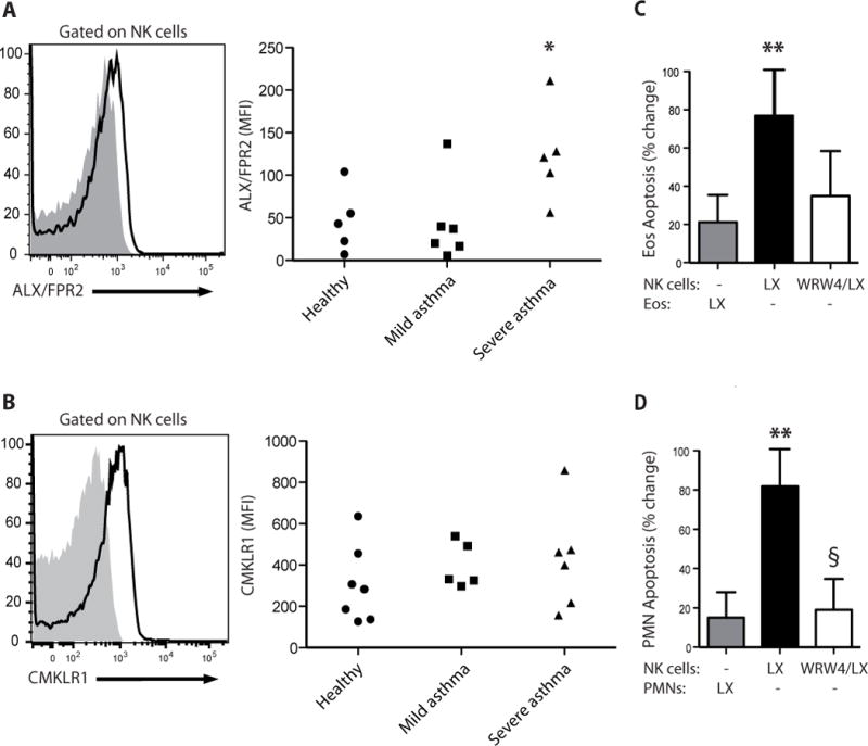
(A) Flow cytometry histograms for anti-ALX (FPRL1) Ab (white) and isotype control (gray) for peripheral blood NK cells (left panel) and MFI for ALX expression on peripheral NK cells in healthy subjects and mild and severe asthmatic subjects (right panel). (B) Flow cytometry histograms for anti-CMKLR1 Ab (white) and isotype control (gray) for peripheral blood NK cells (left panel) and MFI for CMKLR1 expression on peripheral NK cells in healthy subjects and mild and severe asthmatic subjects (right panel). *, P < 0.05, compared with healthy and mild asthmatic subjects (one-way ANOVA). (C, D) Autologous granulocytes and NK cells from healthy subjects were selectively exposed to lipoxin A4 (LX, 100 nM, 15 min, 37°C) in the absence or presence of the ALX/FPR2 antagonist (WRW4, 230nM) prior to co-incubation and the percent change in apoptosis was determined for (C) eosinophils (Eos) and (D) neutrophils (PMN) (see Methods). Results are expressed as mean ± SEM; n≥4; **, P < 0.05, compared with control (one-way ANOVA); §, P < 0.05, compared with NK(LX).