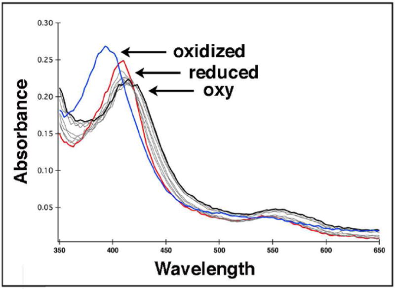
Figure 1.

Stopped flow absorption spectra showing oxy-P450cin complex formation. One syringe contained deoxygenated 20 μM P450cin in 50 mM KPi, pH 7.4, 50 mM KCl with oxygen scrubbers. The other syringe contained only oxygen saturated buffer. Upon mixing, the Soret peak of reduced P450cin (red) immediately shifts from 411 nm to 418 nm (black). Autooxidation to Fe(III) occurs rapidly with a Soret peak shift to 392 nm (blue). The oxidized spectrum was recorded 10 seconds after mixing so the lifetime of the oxy-complex is extremely short.
