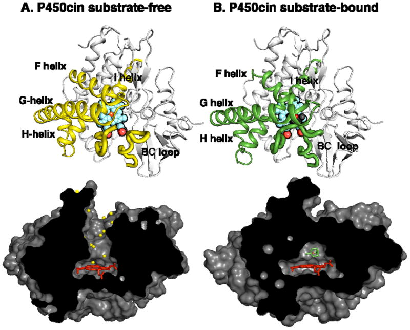
Figure 3.

(A) The P450cin substrate-free and (B) -bound structures. The cartoon representations are viewed directly above the heme plane with regions involved in movement colored yellow for substrate-free and green for substrate-bound. The surface representations are viewed along the heme edge. In the substrate-free structure, the substrate access channel opens up and is lined with solvent (yellow spheres).
