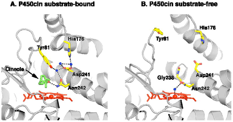
Figure 5.

(A) The substrate bound ferric P450cin structure is depicted with active site waters colored as blue spheres and key residues illustrated in yellow. Black dashes represent hydrogen bonding and coordination interactions. (B) The open substrate-free structure with all solvent removed except for the water coordinated to the heme iron.
