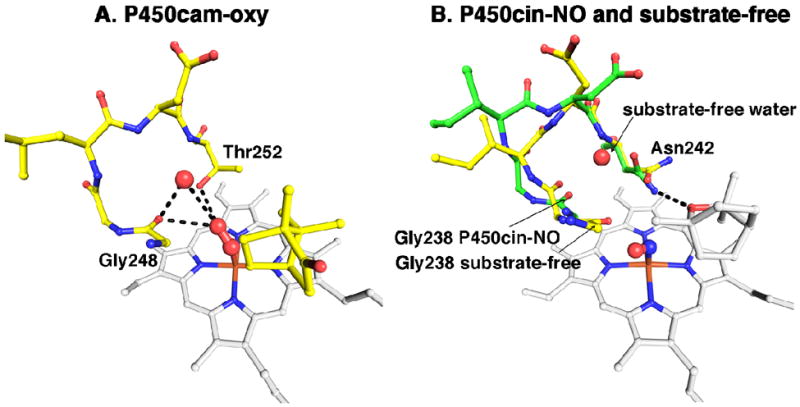
Figure 8.

(A) The P450cam oxy complex active site is colored yellow and the heme colored white. The oxygens and water are represented by red spheres. Hydrogen bonds are drawn as black dashes. (B) NO-P450cin (green) is overlaid with substrate-free P450cin (yellow). Both the heme and substrate are colored white. The NO complex is represented as red and blue spheres just above the heme iron. The water coordinating the heme iron in the substrate-free structure is not shown.
