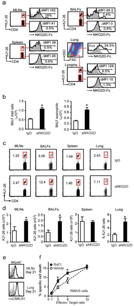
Figure 6. Antigen specific T cells and eosinophils express NKG2D ligands for clearance from inflamed lung.
a Expression of NKG2D ligands on KJ1-26+ and KJ1-26− CD4+ T cells from MLNs, BALFs, spleen and lung eosinophils (Eos) was determined with NKG2D-Fc fusion protein. Histograms show secondary alone (gray) and expression of ligands (black). Inserts show the delta (Δ) median fluorescence intensity (MFI-MFI control) and the percentage of cells positive for NKG2D ligands. Mice were depleted of endogenous NK cells with aGM1 and reconstituted with donor NK cells that were exposed ex vivo to anti-NKG2D (aNKG2D). b BALF total cells and eosinophils were enumerated after aNKG2D or IgG control antibody. c Flow cytometry plots from CD4+ T cells in MLNs, BALFs, spleen and lung. Inserts show percentages of CD4+ KJ1-26+ T cells. d The number of CD4+ KJ1-26+ T cells in MLNs, BALFs, spleen and lung (percentage) after aNKG2D or control antibody. e Representative histograms show expression of the RvE1 receptor CMKLR1 on NK cells (NKp46+ CD3−) from MLNs and lung (day 21). f NK cell cytotoxicity towards RMA/S target cells was determined in the presence of RvE1 (10 nM) or vehicle control * P <0.05 (vehicle), § P <0.05 (RvE1). Data (mean ± s.e.m) are representative of more than 3 independent experiments with n≥4 BALB/cj mice in each group.

