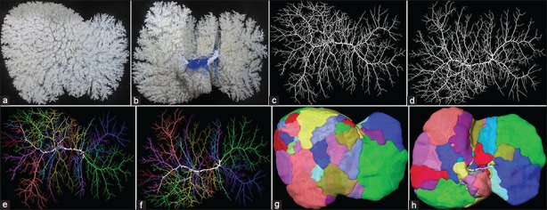
Figure 1.

Portal venous territories in a human liver. All figures are from the same liver, thus excluding any anatomical variation in the branching pattern. (a) Front view and (b) the posterior (also called inferior) view of the native portal venous corrosion cast; (c) Front view and (d) the posterior view of the portal venous branching pattern as reconstructed from CT images. The liver investigated had 24 second-order branches; (e) Front view and (f) the posterior view reveal the portal venous branching pattern with all second-order branches of the liver and the major third-order branches to the right hemiliver marked by different colors; (g) Front view and (h) the posterior view of the corresponding second and third-order territories.
