
Fig 7.
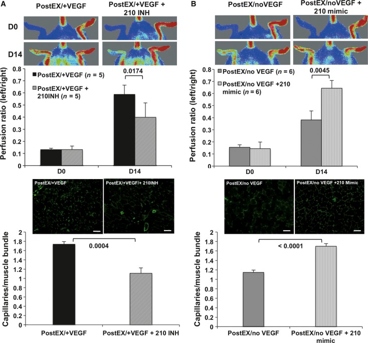
MiR-210 promotes tissue perfusion and capillary density by expanded CD34+ cells in mice ischaemic hindlimb. Expanded cells were transfected for 48 hrs with either miR-210 inhibitor or mimic on day 5. After washing, 2.5 × 10⁴ cells were injected into ischaemic limb. MiR-210 inhibition abrogated tissue re-perfusion (A, upper panel) and capillary density (A, lower panel) in postEX/+VEGF group, whereas mimic significantly improved tissue re-perfusion (B, upper panel) and capillary density (B, lower panel) in postEX/noVEGF. Representative images of tissue perfusion and capillary density in calf muscle assessed by CD31staining (×200; scale bar = 200 μm) are shown in the right upper and lower panels, respectively.