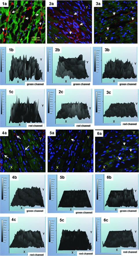
Fig 5.

Analysis of Immunofluorescence profiles of HIF1α and VEGF proteins in sections of rat hearts subjected to ischaemia/reperfusion (I/R) and treatments with γ-tocotrienol, resveratrol or longevinex. Experimentalconditions: Panels (1a-1c) are I/R+vehicle only; Panels (2a-2c) are sham treatment; Panels (3a-3c) are I/R + γ-tocotrienol; Panels (4a-4c) are I/R+ resveratrol; Panels (5a-5c) are I/R+ γ-tocotrienol + resveratrol; Panels (6a-6c) are I/R+ longevinex. In the presented panels multichromatic images of projections of nuclear factor HIF1 (green channel), and VEGF (red channel) are shown in Panels a, while the relative immunofluorescence intensities of HIF1α and VEGF are shown in Panels b, and Panels c respectively. Note: counterstainingof nuclei is presented in blue color. Spatial co-localization of HIF1 and VEGF is indicated with white arrows.
