
Fig 3.
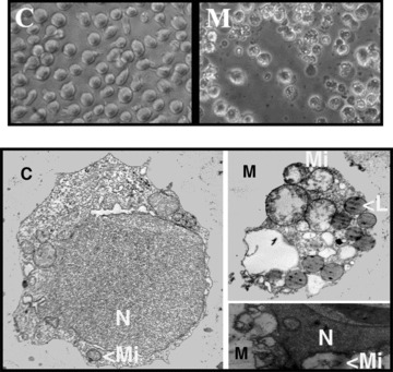
Upper panels: phase contrast microscopy (original magnification × 400) of Jurkat cells cultured for 72 hrs in the presence (M) or absence (C) of 50 μM Minerval. Minerval-treated cells show membrane blebbing and budding and cellular degeneration which was further assessed by electron microscopy. Lower panels: transmission electron microscopy of Jurkat cells incubated in the absence (Control, C, left, original magnification × 5,000) or presence of Minerval (M, right, original magnification × 8,000). The right TEM panels show in detail two apoptotic bodies also observed by phase contrast microscopy in the presence of Minerval. N, nucleus; Mi, mitochondria; L, lipid vesicles.