
Fig 4.
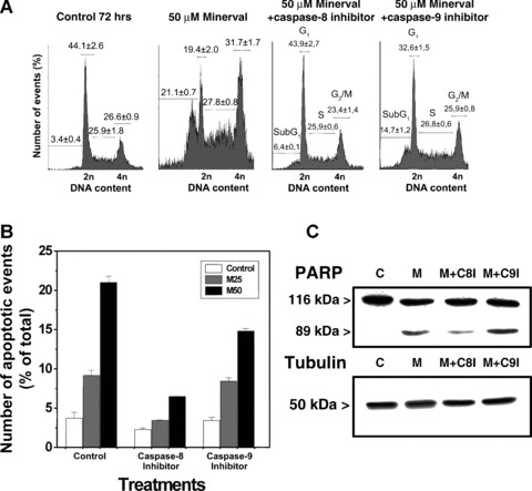
Effect of caspase-8 and caspase-9 inhibition on Minerval-induced apoptosis. (A) DNA content in Minerval-treated cells (50 μM) in the presence of caspase-8 (left) or caspase-9 (right) inhibitors. Basal apoptosis in the presence of 50 μM Minerval was 21.1% of the cells in culture. (B) Jurkat cells were cultured for 72 hrs in the presence or absence (open bars) of 25 (M25, grey bars) or 50 μM (M50, black bars) of Minerval, and in the presence or absence (control) of caspase-8 and caspase-9 inhibitors. C, PARP fragmentation in untreated Jurkat cells (control, C) and in cells treated with 50 μM Minerval in the presence or absence (M) of the caspase-8 inhibitor (M+C8I) or the caspase-9 inhibitor (M+9I). Immunoreactive α-tubulin bands from cells treated as above are also shown.