
Fig 5.
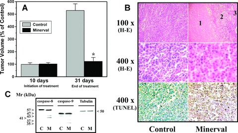
Effect of Minerval treatments on tumour growth in mice. (A) Nude mice were infected with Jurkat cells by s.c. injection on the dorsal surface. Ten days after implantation, tumours were measured and then animals were treated with vehicle (Control, grey bars) or Minerval (black bars) for another 21 consecutive days. The bars correspond to the mean ± SEM values of tumour volume at day 10 (before treatment) and 31 (end of treatment) post-implantation. (B) Histopathological analysis of tumours dissected from vehicle- (Control, left) and Minerval-treated (right) animals. Tumours from control animals showed a homogeneous histological organization, being most cells alive. In tumours from animals treated with Minerval (which were smaller than those from vehicle-treated animals), the greatest region corresponded to that of dead cells (1), followed by a transition area (2) containing dead cells and cells with pycnotic nuclei and a small region (3) in which most cells were alive. The lower micrographs of this panel show that most cells in Minerval-treated animals underwent apoptosis (TUNEL). (C) Immunoblotting detection of caspase-8, caspase-9 and tubulin in tumours from untreated (Control) and Minerval-treated (M) mice.