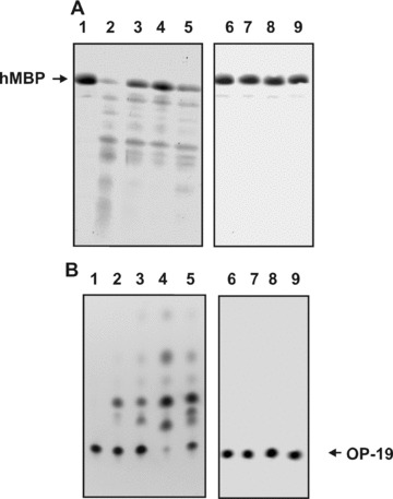
Fig 1.

Examples of hMBP and OP-19 hydrolysis by non-fractionated pIgGs from different MS patients and control IgGs. The hydrolysis of 0.19 mg/ml hMBP (2.5 hrs) in the presence of pIgGs was followed by the decrease in the intensity of Coomassie-stained hMBP band after SDS-PAGE electrophoresis (A), and the hydrolysis of 0.33 mM OP-19 (2.0 hrs), by the decrease in the fluorescence of initial OP-19 spots after TLC (B). The difference in the intensities of these substrates incubated in the absence (lane 1) and in the presence of IgGs from four MS patients (lanes 2–5) was used for the calculations of their RAs. Several control IgGs were used: from a healthy donor (lane 6), a patient with schizophrenia (lane 7), IgGs from MS patients having no affinity for hMBP-Sepharose (lane 8) and IgGs from a healthy patient having affinity for hMPB-Sepharose and purified on this sorbent (lane 9). To quantitatively estimate the protease activity only experiments with the 15–40% conversation of substrate to its product of hydrolysis were used, for example, lane 4 (A) and lines 2 and 3 (B). In this experiment, 0.1 mg/ml pIgGs from eight different patients were used.
