
8.
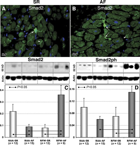
Representative immunofluorescent images of Smad2 labelling in RA appendages in patients in SR (A) and in patients with AF (B). Nuclei are stained blue with DAPI. Arrows denote fibroblast-like cells and arrowheads indicate intercalated disk-like structures. Typical WB and quantitative WB data of Smad2 (C) and Smad2ph (D) in different atrial tissues in patients in SR and in patients with AF.